Client Type
Internal project
Unmet Need
With a recent heightening of accreditation requirements, there is an unmet need for ophthalmic simulators that can present a range of anatomical variations and pathologies to students and professionals in training in ophthalmology and optometry.

Approach
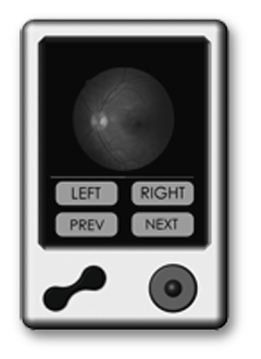
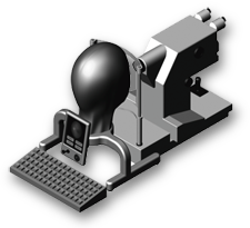
A computer-driven, backlit microdisplay mounted inside a lifelike manikin head provides realistic simulation of the retina. The trainee views the images through any commercial fundus camera or retinal imager.
The “eyes” of the manikin are lenses that direct the view of the camera onto the microdisplay. Images stored in the computer are called by an instructor and displayed in the simulator, evoking trainee responses. Images may be captured using the equipment being used for training and evaluated by the instructor.
Product Features
Simulators eliminate the need for human subjects during training and provide more consistent and repeatable teaching conditions.
Sequences of images may be used for
standardized testing of fundus photography and fluorescein angiography techniques.
Areas of Expertise Provided by OTI
- System conceptualization
- Preliminary optical design and engineering
- Preliminary electronic HW & SW layouts
- SBIR proposal generation
